MED SURG II - EXAM #1 - Rhythm Strip Analysis and Dysrhythmias

Question 1
Contains a rate between 60-100 bpm with a regular rhythm. The QRS is usually normal in shape and duration, P wave is normal and consistent, and always in front of QRS. The PR interval is consistent and between 0.12 and 0.20 seconds, with a PQRS Ratio of 1 to 1
A Normal Sinus Rhythm
Sinus Bradycardia
Sinus Tachycardia
Premature Atrial Complex
Question 2
Often caused by lower metabolic needs such as sleep, athletic training, and hypothyroidism, vagal stimulation such as vomiting, suctioning, severe pain, medications such as calcium channel blockers, or beta-blockers, idiopathic sinus node dysfunction, ICP, and CAD.
Sinus Bradycardia
Premature Atrial Complex
Sinus Tachycardia
Atrial Fibrillation
Question 3
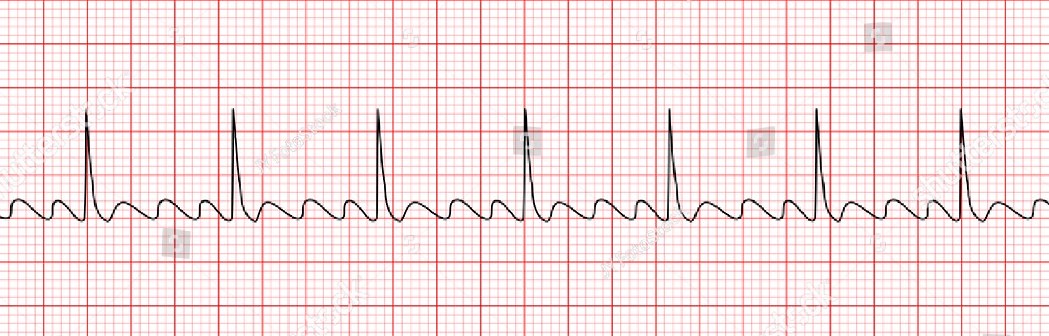
Atrial Fibrillation
Premature Atrial Complex
Atrial Flutter
Sinus Tachycardia
Question 4
Thought to represent repolarization of the Purkinje Fibers. This wave is rare but it sometimes appears in patients with hypokalemia, hypertension, or heart disease. It usuall follows the T wave, and can often be mistaken for a P wave
T Wave
S Wave
U Wave
R Wave
Question 5
May give atropine IV repeated every 3-5 minutes until max dose of 3 mg is given. Rarely, it is unresponsible, but if it is, emergency transcutaneous pacing can be instituted, or medications such as dopamine or epinephrine are given.
Treatment for Atrial Fibrillation
Treatment for Bradycardia
Treatment for Tachycardia
Treatment for Premature Atrial Complex
Question 6
Atrial rate between 250 and 400 bpm, ventricular rate between 75 and 150 bpm. The rhythm is usually regular but can turn irregular in the ventricular valves, the QRS is usually normal but can be abnormal or absent. P wave shows a saw-toothed appearance. PQRS rate is 2 to 1, three to 1, or four to one
Premature Atrial Complex Characteristics
Atrial Fibrillation Characteristics
Atrial Flutter Characteristics
Sinus Tachycardia Characteristics
Question 7
Represents the electrical impulse that starts in the SA node and spreads through the atria (Atrial repolarization). Normally is 2.5 mm or less in height, and 0.11 seconds in duration
U Wave
Q Wave
T Wave
P Wave
Question 8
A normal heart rhythm contains
A P Wave
A QRS Complex
An ST Wave
A T Wave
A U Wave
Question 9
Represents ventricular repolarization, when the cells regain a negative charge, also called the resting state. Usually in the same direction as the QRS, and follows the QRS.
T Wave
Q Wave
U Wave
R Wave
Question 10
Can be caused by physiological or psychological stress such as acute blood loss, anemia, shock, hypervolemia or hypovolemia, heart failure, pain, fever, medications, nicotine, caffeine, drugs, enhanced automaticity of the SA node, and excessive sympathetic tone with a reduced parasympathetic tone that is out of proportion to physiological demands Autonomic dysfunction.
Sinus Bradycardia
Atrial Flutter
Sinus Tachycardia
Premature Atrial Complexes
Question 11
The most common sustained dysrhythmia. It causes an electrophysiologic change in the atrial myocardium and fibrosis. Often caused by atherosclerosis, heart failure, congenital heart disease, COPD, hypothyroidism, and thyrotoxicosis. Some may be asymptomatic but the clinical manifestations include palpitations, dyspnea, and pulmonary edema.
Atrial Flutter
Atrial Fibrillation
Sinus Tachycardia
Sinus Bradycardia
Question 12
Atrial rhythms originate in
SA Node
AV Node
ST Node
SV Node
Question 13
When the atrial ectopic pacer fires at a rate of 250 to 400 bpm, and occurs in a variety of diseases such as rheumatic, coronary, hypertensive, cardiomyopathy, hypoxia, and heart failure. Usually caused by heart failure, tricuspid valve or mitral valve disease, PE, corpulmonale, inferior wall MI, carditis, or digoxin toxicity.
Sinus Bradycardia
Sinus Tachycardia
Atrial Fibrillation
Atrial Flutter
Question 14
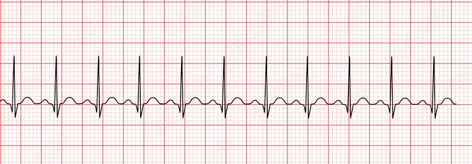
Atrial Fibrillation
Sinus Tachycardia
Premature Atrial Complex
Sinus Bradycardia
Question 15
If the patient is unstable with a ventricular rate of greater than 150 bpm, prepare for immediate cardioversion. if they are stable treatment may include calcium channel blockers, beta blockers, or antidysrhythmics. Anticoagulation may be necessary as there would be pooling of blood in the atria
Premature Atrial Complex Treatment
Atrial Fibrillation Treatment
Atrial Flutter Treatment
Sinus Tachycardia Treatment
Question 16
Vagal manuevers, such as carotid sinus massage, gagging, bearing down in bowel movements, forceful coughing. Administration of adenosine followed by synchronized cardioversion. Beta-blockers, and calcium channel blockers may also be used if the other treatments fail.
Treatment for Sinus Bradycardia
Treatment for Sinus Tachycardia
Treatment for Atrial Flutter
Treatment for Atrial Fibrillation
Question 17
Characterized as tachycardia without hypotension, and by presyncope, palpitations, lightheadedness, weakness, blurred vision, which can occur suddenly with postrual changes.
Inappropriate Sinus Tachycardia
Postural Orthostatic Tachycardia Syndrome
Sinus Tachycardia
Atrial Fibrillation
Question 18
Traces the early part of ventricular repolarization. Normally it is isoelectric, so it is analyzed to identify whether it is above or below the isoelectric line, which may be a sign of cardiac ischemia
TP Segment
QRS Complex
ST Segment
PP Interval
Question 19
If they are infrequent, no treatment is necessary. If there are more than 6 per minute, this is a sign of a worsening condition and may need antidysrhythmics such as procainamide, and quinidine, carotid sinus massage, and reducing caffeine and fixing hypokalemia
Treatments for Atrial Fibrillation
Treatments for Atrial Flutter
Treatments for Premature Atrial Complex
Treatments for Sinus Tachycardia
Question 20
Used to determine atrial rate and rhythm
PP Interval
RR Interval
ST Segment
QRS Complex
Question 21
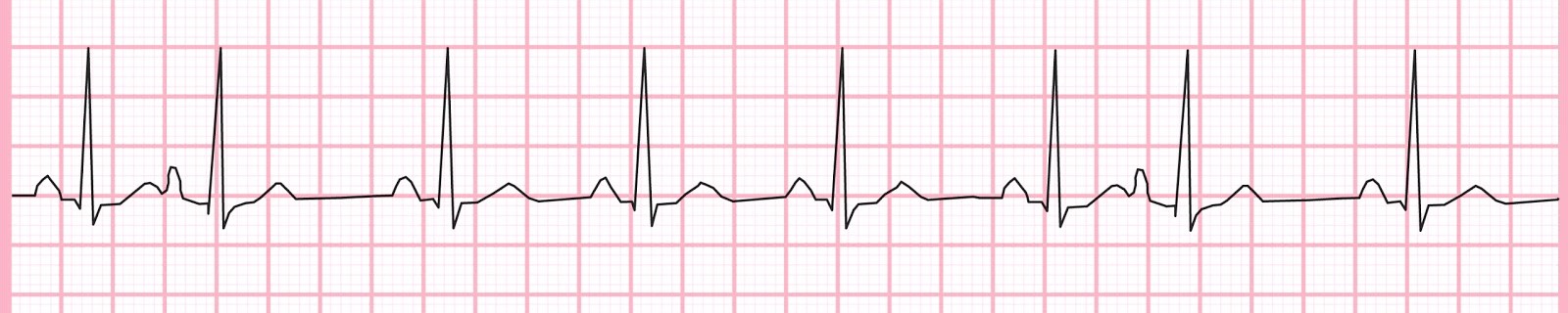
Atrial Flutter
Sinus Bradycardia
Premature Atrial Complex
Sinus Tachycardia
Question 22
Indicates atriventricular conduction time. The time needed for sinus node depolarization, and conduction through the AV node before ventricular depolarization. Usually is 0.12-0.20 seconds
PP Interval
PR Interval
RR Interval
QT Interval
Question 23
Atrial rate is 300 to 600 bpm, and ventricular rate is usually 120 to 200 bpm. The rhythm is highly irregular. QRS is usually normal. There are no discernible P waves, often show as F waves, or snakes in the grass. The PR interval cannot be measured, and the PQRS ratio is many to one
Atrial Fibrillation Characteristics
Atrial Flutter Characteristics
Sinus Bradycardia Characteristics
Sinus Tachycardia Characteristics
Question 24
Often caused by nicotine, alcohol, hypervolemia, anxiety, hypokalemia, pregnancy, or atrial ischemia, injury, or infarction. They are often seen in tachycardia and patients may report palpitations. They are ectopic beats that originate in the atria and are not rhythms, often have an abnormal looking P wave
Premature Atrial Complex
Sinus Tachycardia
Atrial Fibrillation
Sinus Bradycardia
Question 25
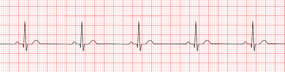
Sinus Tachycardia
Sinus Bradycardia
Premature Atrial Complex
Atrial Fibrillation
Question 26
The rate depends on the underlying rhythm. The rhythm is irregular due to early P waves. The QRS that follows the P wave is usually normal, but may be abnormal or even blocked. The PR interval is shorter than normal but still between 0.12-0.20 seconds. The ratio remains between 1 and 1
Premature Atrial Complex Characteristics
Atrial Fibrillation Characteristics
Atrial Flutter Characteristics
Sinus Tachycardia Characteristics
Question 27
Used to determine ventricular rate and rhythm
PP Interval
PR Interval
RR Interval
SR Interval
Question 28
Enhanced automaticity of the SA node or excessive sympathetic tone with reduced parasympathetic tone that is out of proporation to physiologic demands. Catheter ablaion of the SA node may be used in cases of persistent and unresponsive to other treatments
Inappropriate Sinus Tachycardia
Postural Orthostatic Tachycardia Syndrome
Sinus Tachycardia
Premature Atrial Complex
Question 29
Indicates ventricular depolarization. Usually is less than 0.12 seconds
QRS Complex
ST Segment
PP Interval
PR Interval
Question 30
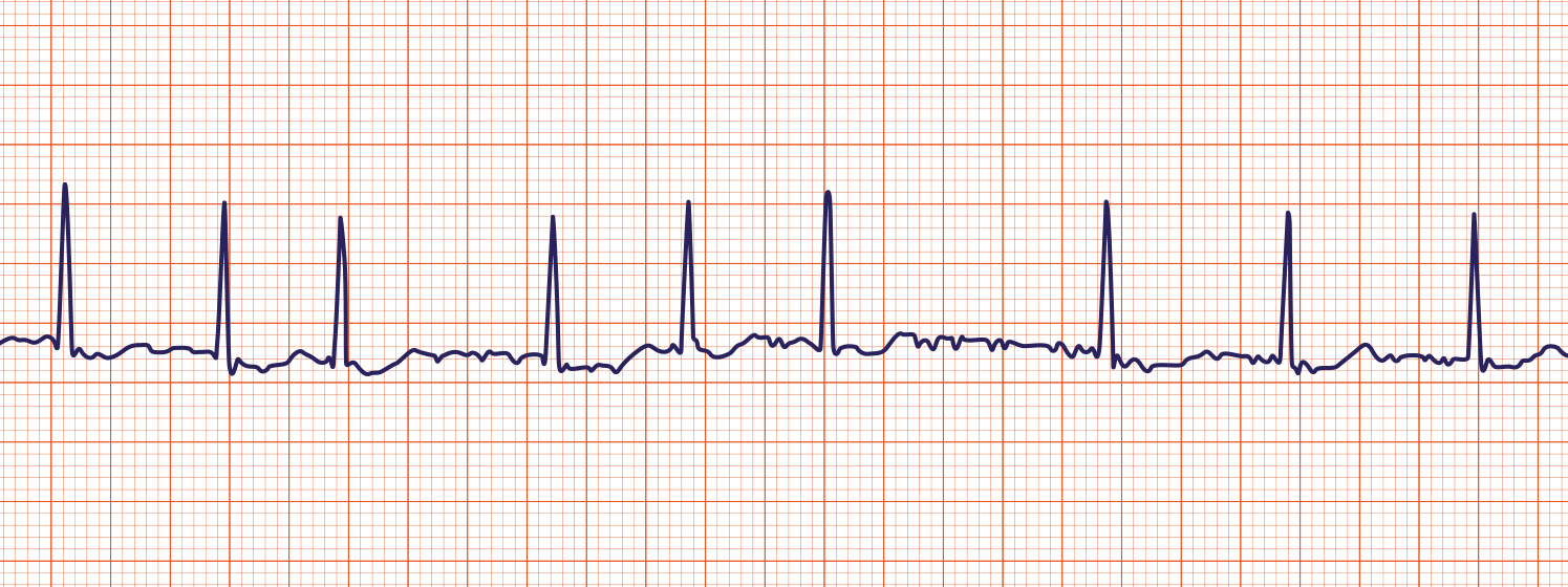
Sinus Bradycardia
Atrial Flutter
Atrial Fibrillation
Sinus Tachycardia
Question 31
While reading a patient's ECG, what would you note to indicate an acute MI?
ST-Segment Inversion
ST-Segment Elevation
T Wave Inversion
T Wave Elevation
An Abnormal Q Wave
Question 32
While assessing a patient, you notice the ECG shows an abnormal Q wave, but other leads are normal. You interpret this as
A valve disorder
Heart failure
An acute MI
An Old MI
An Impending MI